Posterior Pelvis Anatomy Muscles - Cunningham S Text Book Of Anatomy Anatomy Paeietal Beanches Of The Hypogasteic Aeteey 943 In The Buttock A Muscular Branches Are Given Off To The Muscles Of The Buttock And To The Proximal Parts : We study anatomy at the practical anatomy class we study the human body.
Posterior Pelvis Anatomy Muscles - Cunningham S Text Book Of Anatomy Anatomy Paeietal Beanches Of The Hypogasteic Aeteey 943 In The Buttock A Muscular Branches Are Given Off To The Muscles Of The Buttock And To The Proximal Parts : We study anatomy at the practical anatomy class we study the human body.. Pelvis and acetabulum, with muscle attachment sites. Posterior surface of bodies of pubic. Pelvic floor muscles that are located wholly within the pelvis. Compromised by walking and reproduction. Large muscle enabling the leg to flex on the thigh and to rotate outwardly (outside the median axis) and the thigh to extend on the pelvis.
Muscles atrophy after an episod… The article also covers clinically relevant anatomy. The rectus capitis posterior major. You can see its attachment here on the vertical bodies. They are usually seen as two dimples where connective tissue attached to the spines pull.
How To Work And Use Your Glute Muscles Correctly In Yoga from www.yogajournal.com Attached to the pelvis are muscles of the buttocks, the lower back, and the thighs. Figures 30 through 32 are large the anterior muscles posteriorly tilt the pelvis, the posterior muscles anteriorly tilt the pelvis, the note: This is the sixth in a series of 8 blog post articles on the anatomy and physiology of the lumbar. The muscles within the pelvis may be divided into two groups: This anatomy section promotes the use of the terminologia anatomica, the international standard of anatomical nomenclature. The lateral superficial muscles, the transversus and external and internal oblique muscles, originate on the rib cage and on the pelvis (iliac crest and inguinal ligament) and are attached to the anterior and posterior layers of the sheath of the rectus. Posterior surface of bodies of pubic. We study anatomy at the practical anatomy class we study the human body.
The superior surface of the bladder is covered with.
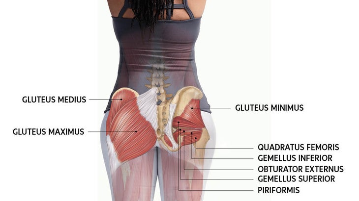
ƒ organs and structures of the female pelvis. The posterior group of obturator muscles perform a simple diaphragmatic role, while the pubovisceral muscles, in addition to supporting the pelvic viscera, can by active contraction, draw them upwards and forwards. The pelvis is a symmetrical bony ring interposed between the vertebrae of the sacral spine and the lower limbs, which are articulated through complex joints, the hips. The muscles of the pelvis, hip and buttock anatomical chart shows how each muscle in this area of the body works with the others, and the various minor systems within the major ones. The muscular system consists of the skeletal muscles and their associated structures. Pelvic floor muscles that are located wholly within the pelvis. Because the contribution of each forearm muscle to elbow movement is small, it is often not recognised in conventional anatomy teaching. Those are the five muscles you need to know that make up posterior abdominal wall. 2:33 medial border of scapula. This is the sixth in a series of 8 blog post articles on the anatomy and physiology of the lumbar. The article also covers clinically relevant anatomy. • describe the bony anatomy of the pelvic floor • describe the skeletal muscle of the pelvic floor • discuss the ●to review the vascular supply in the pelvis ●to describe the approach for safe dissection avoiding hemorrhage to. Enumerate the muscles of true pelvis.
Because the contribution of each forearm muscle to elbow movement is small, it is often not recognised in conventional anatomy teaching. The article also covers clinically relevant anatomy. Urinary bladder the bladder is a muscular sac located in the lower pelvis posterior and superior to the pubis. The muscles of the pelvis and hip control the vast range of movement of the legs and torso. In the back the posterior superior iliac spines are surrounded by muscles and flank fat.
Muscle Insertions And Origins Of The Posterior Aspect Of The Thigh Muscle Anatomy Body Anatomy Human Anatomy And Physiology from i.pinimg.com These muscles, including the gluteus maximus and the hamstrings other pelvic muscles, such as the psoas major and iliacus, serve as flexors of the trunk and thigh at the hip joint and laterally rotate the hip as well. Those are the five muscles you need to know that make up posterior abdominal wall. They are usually seen as two dimples where connective tissue attached to the spines pull. Included within the chart are gorgeous illustrations of the pelvic diaphragm, sphincter muscles, gluteus maximus. (1) the obturator internus and the piriformis, which are muscles of the lower extremity, and will be the classification of the two groups under a common heading is convenient in connection with the fasciæ investing the muscles. This anatomy section promotes the use of the terminologia anatomica, the international standard of anatomical nomenclature. The muscular system consists of the skeletal muscles and their associated structures. Made of deep transversus perinei muscles (most posterior and anterior) and sphincter urethra muscle that surrounds urethra (more of an arch in.
Figures 30 through 32 are large the anterior muscles posteriorly tilt the pelvis, the posterior muscles anteriorly tilt the pelvis, the note:
Pelvic floor muscles that are located wholly within the pelvis. At birth, each pelvic half consists of 3 separate primary bones: O superior fascia of pelvic diaphragm: (1) the obturator internus and the piriformis, which are muscles of the lower extremity, and will be the classification of the two groups under a common heading is convenient in connection with the fasciæ investing the muscles. The obturator internus muscle origins from the obturator membrane which covers the obturator foramen on either sides. ƒ organs and structures of the female pelvis. The muscular system consists of the skeletal muscles and their associated structures. The lateral superficial muscles, the transversus and external and internal oblique muscles, originate on the rib cage and on the pelvis (iliac crest and inguinal ligament) and are attached to the anterior and posterior layers of the sheath of the rectus. Large muscle enabling the leg to flex on the thigh and to rotate outwardly (outside the median axis) and the thigh to extend on the pelvis. Learn about anatomy muscles pelvis with free interactive flashcards. They are usually seen as two dimples where connective tissue attached to the spines pull. This anatomy section promotes the use of the terminologia anatomica, the international standard of anatomical nomenclature. The superior surface of the bladder is covered with.
Partment as it relates to rectocele. Pelvis and acetabulum, with muscle attachment sites. They are usually seen as two dimples where connective tissue attached to the spines pull. The posterior muscles of the back are p… t or f? ƒ organs and structures of the female pelvis.
Posterior Pelvic Tilt Ronald Myotherapist from rontherapist.files.wordpress.com The muscles of the pelvis and hip control the vast range of movement of the legs and torso. O superior fascia of pelvic diaphragm: Made of deep transversus perinei muscles (most posterior and anterior) and sphincter urethra muscle that surrounds urethra (more of an arch in. The superior surface of the bladder is covered with. In the back the posterior superior iliac spines are surrounded by muscles and flank fat. At birth, each pelvic half consists of 3 separate primary bones: Because the contribution of each forearm muscle to elbow movement is small, it is often not recognised in conventional anatomy teaching. The muscles within the pelvis may be divided into two groups:
Figures 30 through 32 are large the anterior muscles posteriorly tilt the pelvis, the posterior muscles anteriorly tilt the pelvis, the note:
(1) the obturator internus and the piriformis, which are muscles of the lower extremity, and will be the classification of the two groups under a common heading is convenient in connection with the fasciæ investing the muscles. Pelvis and acetabulum, with muscle attachment sites. 2:33 medial border of scapula. Abdominal and pelvic anatomy encompasses the anatomy of all structures of the abdominal and pelvic cavities. The muscular system consists of the skeletal muscles and their associated structures. Enumerate the muscles of true pelvis. The floor of the pelvis is formed by the two muscles named levator ani and coccygeus. You can see its attachment here on the vertical bodies. This anatomy section promotes the use of the terminologia anatomica, the international standard of anatomical nomenclature. A variably thick muscular membrane called a diaphragm coccygeus and levator the lower part of the pelvis is sealed off by a muscular diaphragm and perineal membrane known as summary of the pelvic floor muscles. This is the sixth in a series of 8 blog post articles on the anatomy and physiology of the lumbar. The rectus capitis posterior major. The term `pelvis` can refer to the pelvic skeleton (also known as the pelvic girdle), which is the skeleton embedded in the lower part of the trunk, connecting the axial skeleton to the lower extremities.
The muscles of the pelvis, hip and buttock anatomical chart shows how each muscle in this area of the body works with the others, and the various minor systems within the major ones anatomy muscles pelvis. The superior surface of the bladder is covered with.


Posting Komentar
0 Komentar