Leg Muscle Diagram Side / Hamstring Muscles And Your Back Pain - The achilles tendon is also located in the lower leg.. Vector illustration informative medical scheme. Observe the leg muscle diagram posted above and notice that there are many parts in the muscles. Leg muscles diagram / drawings: This image shows the muscles of our body and displays them on both male and female diagram showing: Together, these muscles straighten your knee, stabilize your knee joint, assist in flexing your hip (drawing your knee towards your chest), and help absorb force when you land after jumping or leaping.
The biceps femoris is a muscle of the posterior thigh composed of a long head and a short head. Leg muscle anatomical structure, labeled front, side and back view diagrams. The peroneus longus and brevis both have their origins on the fibula and they both pass behind the lateral malleolus where their tendons pass under the peroneal retinacula. Muscle anatomy interactive 12 photos of the muscle anatomy interactive human muscle anatomy interactive, interactive muscle anatomy games, muscle anatomy interactive, muscle anatomy interactive quiz, shoulder muscle anatomy interactive, human muscles, human muscle anatomy interactive, interactive muscle anatomy games. The largest muscle masses in the leg are present in the thigh and the calf.
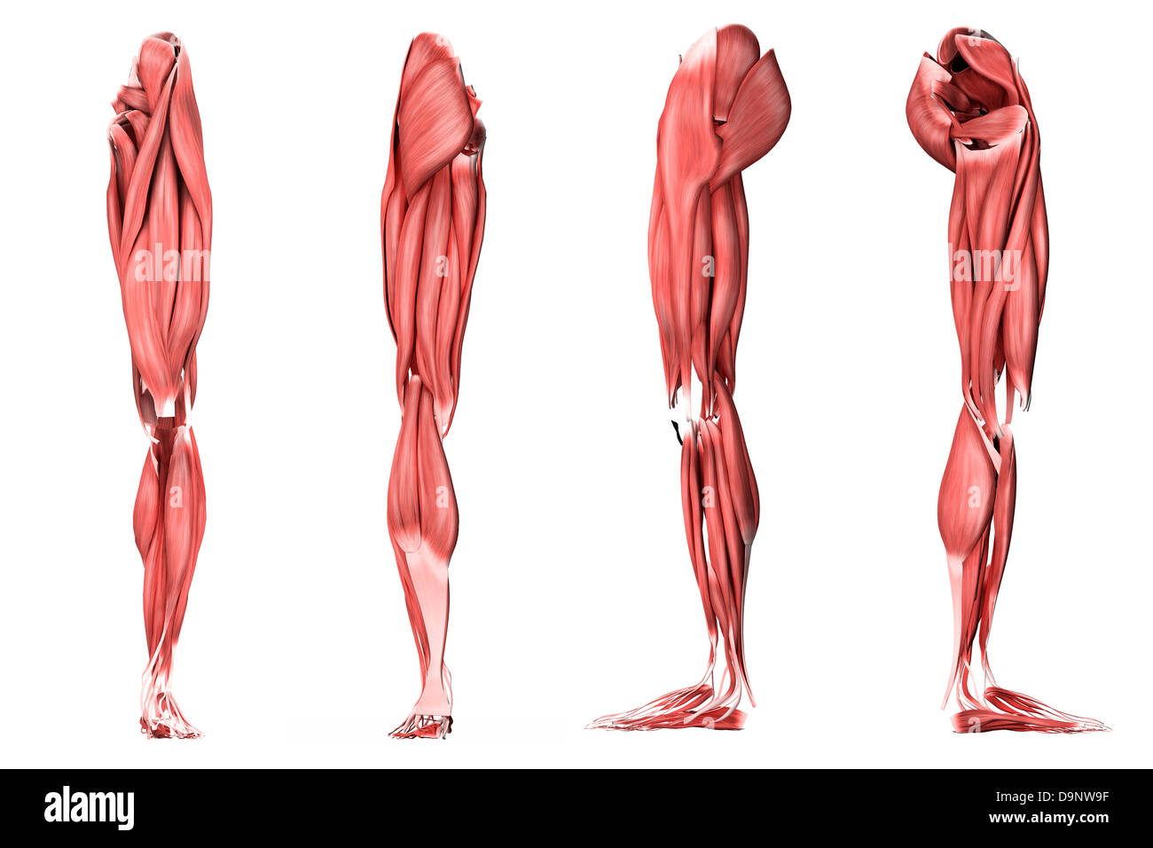
Medical Illustration Of Human Leg Muscles Four Side Views Stock Photo Alamy from c8.alamy.com It begins in the lower back and runs down to the. The major nerve of the leg is the sciatic nerve. This muscle is located on the side of the leg and is actually on the other side of the large toe which it is responsible for moving. Leg muscle anatomical structure, labeled front, side, and back view diagrams. A muscle along the outside of the leg that bends the foot out at the ankle. Nerves in the leg send messages to the brain, including indications of heat, pain, and movement. The achilles tendon is also located in the lower leg. Detailed anterior, lateral and posterior views.men sports fitness training.
Leg muscle anatomical structure, labeled front, side, and back view diagrams.
Below the rectus femoris and largely hidden by it is the vastus intermedius. The muscles in the front allow for. These muscles are found on the front and back sides of the lower leg. The fibularis longus originates from the head and upper lateral surface of the fibula, runs in a bony groove along the bottom of the foot to attach on the other side at the base of the first metatarsal and the neighboring medial cunieform bone, and acts to evert the. There are many muscles located in the lower leg, but there are three that are particularly well known—the gastrocnemius and the soleus, which are the most powerful muscles in the lower leg, and the anterior tibialis. Extensor muscles of the hand 6. The largest muscle masses in the leg are present in the thigh and the calf. Editable vector illustrator cc file (editable live text)editable vector eps 10 filehigh resolution jpg filelove and respect from. Because the leg has many different muscles, it is vulnerable to several different types of muscle strains. Your leg muscles are some of the hardest working muscles in your body. Each of these major nerves further divides into many smaller nerve branches to stimulate individual muscles and sense touch, pain, warmth, and cold in the skin. Vector illustration informative medical scheme. Homework for lasalle college of the arts:
Vector illustration informative medical scheme. The major nerve of the leg is the sciatic nerve. The thigh (proximal lower limb) muscles are arranged into three compartments : The muscles in the front allow for. Muscle anatomy interactive 12 photos of the muscle anatomy interactive human muscle anatomy interactive, interactive muscle anatomy games, muscle anatomy interactive, muscle anatomy interactive quiz, shoulder muscle anatomy interactive, human muscles, human muscle anatomy interactive, interactive muscle anatomy games.
1 from Extensor muscles of the hand 6. The biceps femoris is a muscle of the posterior thigh composed of a long head and a short head. The following diagram illustrates the actions of the terms adduction, abduction, flexion and extension at the different joints. Sketching the leg side view / muscles of the anterior compartment of the leg. The muscles of the leg anatomy chart shows in every possible view the way that the muscles and other pieces of the leg work together in motion. The peroneus longus and brevis both have their origins on the fibula and they both pass behind the lateral malleolus where their tendons pass under the peroneal retinacula. Vector illustration informative medical scheme. Related posts of lower leg muscles diagram muscle anatomy interactive.
Two muscles on the lateral side of the leg form the peroneal group.
The short head originates from the lateral lip of linea aspera and. Observe the leg muscle diagram posted above and notice that there are many parts in the muscles. The major nerve of the leg is the sciatic nerve. The lower leg lies between the knee and the ankle. Medial compartment, also known as adductor compartment; Together with the upper leg, it forms the lower extremity. The muscles of the leg anatomy chart shows in every possible view the way that the muscles and other pieces of the leg work together in motion. Because the leg has many different muscles, it is vulnerable to several different types of muscle strains. The thigh (proximal lower limb) muscles are arranged into three compartments : Detailed anterior, lateral and posterior views.men sports fitness training. Some of the more common ones are: The achilles tendon is also located in the lower leg. A muscle strain is a stretch or tear of muscle fibers.
The achilles tendon is also located in the lower leg. The fibularis longus originates from the head and upper lateral surface of the fibula, runs in a bony groove along the bottom of the foot to attach on the other side at the base of the first metatarsal and the neighboring medial cunieform bone, and acts to evert the. Nerves in the leg send messages to the brain, including indications of heat, pain, and movement. Related posts of lower leg muscles diagram muscle anatomy interactive. A muscle strain is a stretch or tear of muscle fibers.
Leg Definition Bones Muscles Facts Britannica from cdn.britannica.com The short head originates from the lateral lip of linea aspera and. Medial compartment, also known as adductor compartment; Vector illustration informative medical scheme. Detailed anterior, lateral and posterior views.men sports fitness training. 849 x 989 jpeg 81 кб. The popliteus works on the knee while the other three are associated with the foot and ankle. Leg muscle anatomical structure, labeled front, side and back view diagrams. The femoral, saphenous, obturator, and lateral femoral cutaneous nerves all extend from the lumbar plexus into the muscles and skin of the thigh and leg.
The lower leg is a major anatomical part of the skeletal system.
Gain, not the most accurate reference but i tried. Two muscles on the lateral side of the leg form the peroneal group. Homework for lasalle college of the arts: Leg muscle anatomical structure, labeled front, side, and back view diagrams. Below the rectus femoris and largely hidden by it is the vastus intermedius. 849 x 989 jpeg 81 кб. Vector illustration informative medical scheme. The lower leg is a major anatomical part of the skeletal system. Leg muscle anatomical structure, labeled front, side and back view diagrams. Leg muscle anatomical structure, labeled front, side and back view diagrams. Because the leg has many different muscles, it is vulnerable to several different types of muscle strains. The major nerve of the leg is the sciatic nerve. Vector illustration informative medical scheme.
Together, these muscles straighten your knee, stabilize your knee joint, assist in flexing your hip (drawing your knee towards your chest), and help absorb force when you land after jumping or leaping leg muscle diagram. Posterior compartment, also known as the flexor compartment;


Posting Komentar
0 Komentar